


TECHNOLOGY
We offer you a wide range of high quality models of bone for workshops, training and display modes for presentation of implant products.
All of our products can be adapted to your requirements and custom made products can be produced according to your suggestions.
 Plastic cortical shell. The plastic cortical shell models are made of a rigid plastic shell with cancellous material in the proximal and distal ends. The bone includes the intramedullary canal 8 to 12 mm diameter, materials version A.
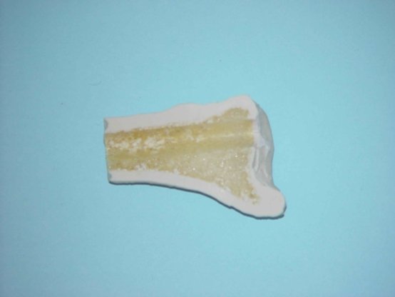 Foam cortical shell. The foam cortical shell models are made of a rigid foam shell with cancellous material in the distal and proximal ends. Also include the intramedullary canal 8 to 12 mm diameter. These models could be cut and drilled ea sier the n the plastic cortical shell models and are most commonly used for total joint replacement, internal fixation and any other orthopedic procedure where a medium cost bone is desired, materials version B.

Solid foam. The solid foam bone models are made of a rigid foam throughout. The bones include the intramedullary canal 8 to 12 mm diameter. The product is most commonly used for external fixation and some limited total joint replacement and internal fixations. It is economical bone for most applications, material version C.
The adaptation of technology towards achievement the optimal real bone parameters
.jpg)
Fig. 5. Cortical Shell, osteocytes, Haversian canal, lamellae of bone
The human bone consists of organic and mineral matter. A protein substance called ossein is the organic component of the bone. It is composed mainly of collagen fibers. The mineral part of the bone consists in 65% of hydroxyapatites. These are complex compounds of calcium carbonate and calcium phosphate with small admixtures of sodium, magnesium, potassium, chlorine and fluorine.

.BMP)
.BMP)
Figure 6.
Figure 7.
.BMP)
.BMP)
Figure 8. Figure 9.
.BMP)
.BMP)
Figure 10. Figure 11.
.BMP)
Figure 12.


Articles about the use of our products All of our products can be adapted to your requirements and custom made products can be produced according to your suggestions.
Plastic cortical shell. The plastic cortical shell models are made of a rigid plastic shell with cancellous material in the proximal and distal ends. The bone includes the intramedullary canal 8 to 12 mm diameter, materials version A. Foam cortical shell. The foam cortical shell models are made of a rigid foam shell with cancellous material in the distal and proximal ends. Also include the intramedullary canal 8 to 12 mm diameter. These models could be cut and drilled ea sier the n the plastic cortical shell models and are most commonly used for total joint replacement, internal fixation and any other orthopedic procedure where a medium cost bone is desired, materials version B.Solid foam. The solid foam bone models are made of a rigid foam throughout. The bones include the intramedullary canal 8 to 12 mm diameter. The product is most commonly used for external fixation and some limited total joint replacement and internal fixations. It is economical bone for most applications, material version C.
The adaptation of technology towards achievement the optimal real bone parameters
The core of the bone is built of a compact substance, which consists of lamellae of bone and osteocytes.

Fig. Structure of bone
Fig. Structure of bone
Fig. 5. Cortical Shell, osteocytes, Haversian canal, lamellae of bone
The human bone consists of organic and mineral matter. A protein substance called ossein is the organic component of the bone. It is composed mainly of collagen fibers. The mineral part of the bone consists in 65% of hydroxyapatites. These are complex compounds of calcium carbonate and calcium phosphate with small admixtures of sodium, magnesium, potassium, chlorine and fluorine.
The internal cancellous structure is made of bone trabeculae of different shapes. In the compact bone, the cells are stacked together with lamellae of bone in an orderly manner around the artery in the Haversian canal. In the cancellous bone one can find different types of lamellae of bone forming a network which houses the marrow cavities. A typical bone structure in an enlarged view is shown on the following picture.
Figure 4 The cancellous structure
A combination of these structures gives the bone the following properties:
- plasticity, which is modeled according to the Wolf - Delpech principle,
- flexibility connected with the properties of collagen that enable the bone to return to its original shape after the stimulus has ceased,
- phase deformation consisting in rhythmic changes of the shape according to the theory of Sutherland.
Formulating the chemical composition and structure of the composite material we make the models for training, we put the emphasis on the accurate reproduction of the properties of the natural bone. For this purpose we examined the bone in vide range of tests. The same technique was applied in the examination of models made of composite materials.
The internal cancellous structure can be reconstructed in two ways:
- Using a closed-cell material, sufficient to reconstruct bone parameters expected by the surgeons performing implantation procedures,
- Using an open-cell material, which may also be used in other medical procedures.
Open-cell material is characterized by absorbability exceeding 90% and is used primarily for the production of bone models intended for trainings consisting in injection of pharmacological drugs or bone cement into the bone. This cement is injected into the fractured bone to stabilize the fracture.
The materials used in our production pass different test, using a variety of research methods, allowing one to assess their suitability for the production of models intended for medical training in both orthopedics and dentistry. Here are some photos taken with a scanning electron microscope, manufactured by Canon. The samples were vacuum covered with gold before the test, to prevent any destruction.
The SEM pictures taken at a closed-cell material show a characteristic structure of the cancellous bone. The only element our material differs from the natural bone is a thin film that separates the adjacent cells. The film stiffens the cancellous structure of the model and replaces the bone marrow cavity and collagen filling the spaces between the wall structure made of hydroxyapatites.
Figure 6.
The structure of the material used by us for the manufacture of the cancellous structure can be seen particularly well on an image with a three-time higher magnification. One can see a film which separates the adjacent cells of the cancellous structure.
Figure 7.
The structure of the open-cell material looks slightly different (see the images below). One can see clear crakes in the construction of the walls separating particular cells. The intercellular spaces are not fully open, but the breaks are sufficient to ensure a high absorbability of this structure. The residual intercellular membranes of the material (in the absence of the organic fill present in the natural bone) stiffen the structure. This solution prevents a spontaneous collapsation of the material and destruction of its structure.
Figure 8. Figure 9.
Other materials were used to reproduce a compact bone normally made of lamellae of bone and osceocytes and the Haversian canals existing between them. Depending on the expected hardness of the bones from D2 to D4, a different structure of the material is applied. A structure of materials reconstructing the compact part can be seen on SEM images.
The typical structure of a material with hardness equivalent to the bone hardness D4 is shown in the following images
Figure 10. Figure 11.
The structure of the material with a hardness of D2 is visible on the following image
Figure 12.
In case of a compact bone, the main parameters which determine a suitability in the training of orthopedic and dental implantation, is the reconstruction of the bone hardness and its typical sensitivity to cutting and drilling. The color is yet another element, but only of aesthetic value.
Other issues are the mechanical parameters of the bone models. They are important for another group of medical models intended for biomechanical research. In this group, the fourth generation bone models are best known on the market. They guarantee a faithful reproduction of the mechanical properties of the bone (Young's modulus as well as compression and yield strengths); the properties related to cutting and drilling being insignificant.
The main features of bones used during the trainings include:
• Diversified structure present at the cross section,
• Mechanical properties ensuring a similarity during the cutting and drilling processes (including formation of typical bone shavings used during the bone reconstruction),
• Characteristic CT image,
• Stable non-irritating odor and easy storage and transportation.
CT IMAGES
Owing to the application of composite materials with different structure and hardness, our bone models perfectly imitate a two-layer structure of natural bones. The painstaking trials and tests resulted in a selection of "B" version material as the one which resembles to the most extent the natural human bone. The "A" version is used mainly for presentations during exhibitions, and the "C" version is the economical one. Imitation of the bone material behaves similarly to the natural bone during drilling and cutting processes.


An additional feature of our bones, often important during the trainings, is a distinctive X-ray image resembling that of the human bones. Samples of the composite material were tested with the use of a CT scanner. The test results in the form of X-ray images, were shown for comparison in such a way that on the left side one can see the image of the natural human bone and on the right side there is the composite bone. Below you can see a few cross-sectional images of bones. Places where the cross-sections were made are designated with lines marked on the horizontal projection of the bones. CT images with the cross-sections show a very strong resemblance of composite bones to the natural human ones.
Owing to the application of composite materials with different structure and hardness, our bone models perfectly imitate a two-layer structure of natural bones. The painstaking trials and tests resulted in a selection of "B" version material as the one which resembles to the most extent the natural human bone. The "A" version is used mainly for presentations during exhibitions, and the "C" version is the economical one. Imitation of the bone material behaves similarly to the natural bone during drilling and cutting processes.
An additional feature of our bones, often important during the trainings, is a distinctive X-ray image resembling that of the human bones. Samples of the composite material were tested with the use of a CT scanner. The test results in the form of X-ray images, were shown for comparison in such a way that on the left side one can see the image of the natural human bone and on the right side there is the composite bone. Below you can see a few cross-sectional images of bones. Places where the cross-sections were made are designated with lines marked on the horizontal projection of the bones. CT images with the cross-sections show a very strong resemblance of composite bones to the natural human ones.
Natural bone.
Composite bone.
Cross section A-A
Cross section B-B
Cross section C-C
We invite you to see our dental models offer.
We can produce for your own OEM models.
Contact:
PROMEDICUS
PROMEDICUS sp. z o.o.
43-190 Mikołów
ul. Brzoskwiniowa 11
tel +48 32 22 62 530
tel/fax +48 32 73 82 642
promedicus@promedicus.com.pl
www.promedicus.co.uk
PROMEDICUS sp. z o.o.
43-190 Mikołów
ul. Brzoskwiniowa 11
tel +48 32 22 62 530
tel/fax +48 32 73 82 642
promedicus@promedicus.com.pl
www.promedicus.co.uk
